Try out: Click a sample image and see Dr. Watson DDS/DMD's interpretion
| On the Cheek | On the Lips | ||||
|---|---|---|---|---|---|
|
|
|

|

|
|
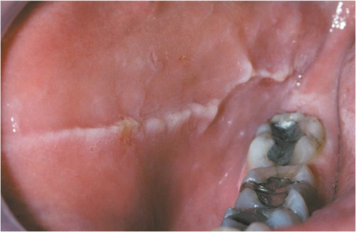
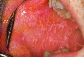
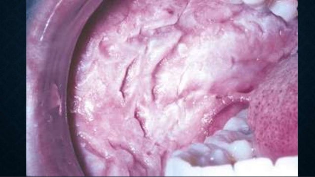
| Linea Alba | Lichen Planus | White Sponge
Nevus |
Herpes
Labialis |
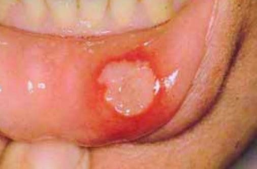
Mucocele | Aphthous
Stomatitis |
| On the Palate or Floor of the mouth | On the Tongue | ||||
|

|
|

|
|

|
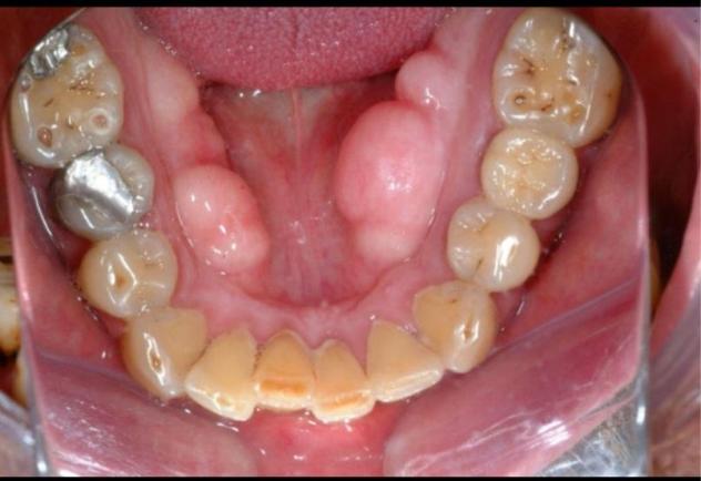
| Torus
Mandibularis |
Torus
Palatinus |
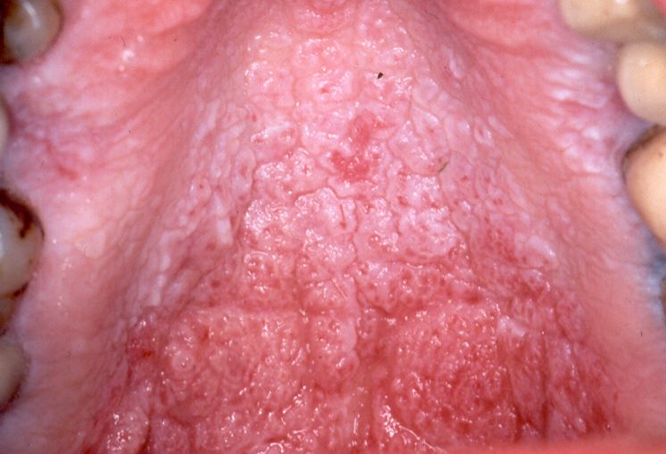
Nicotine
Stomatitis |
Geographic
Tongue |
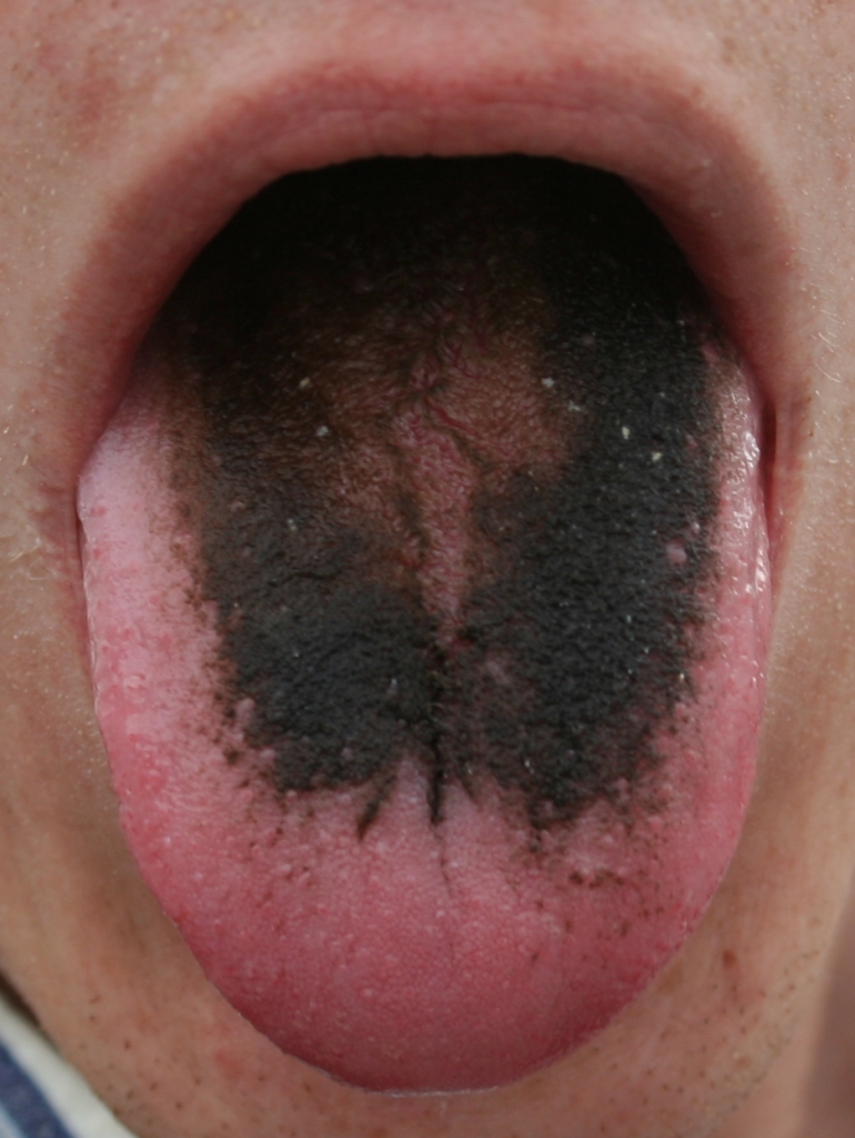
Hairy
Tongue |
Median Rhomboid
Glossitis |
| On the Gingiva or Vestibule area | Or, paste an URL to an intraoral image in JPG or PNG below. | ||||

|
|

|
|||
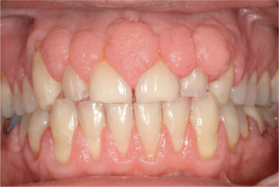
| Necrotizing Ulcerative
Gingivitis & Periodotitis |
Gingival Hyperplasia | Epulis Fissuratum | |||

Dr. Watson DDS/DMD is analyzing the image. Please wait...
Thank you!
Why only those 15 intraoral or clinical findings?
There are many other intraoral lesions and clinical findings in the oral cavity. Why are the only fifteen chosen?
My initial goal for this project is not to develop an AI dentist or oral pathologist which can replace human doctors. But rather, as an oral healthcare professional, I wanted to evaluate IBM Watson to see whether it is selective and sensitive enough to do a doctor's tasks and also to demonstrate how dentistry can benefit from the artificial intelligence and machine learning technology.
Those 15 clinical findings and oral lesions were particularly selected in order to test whether AI can recognize subtle differences in physical and topological appearances despite their common sites of onset in the oral cavity.
For example, linea alba, lichen planus and white sponge nevus occur in the same location, which is the inside of cheeks. Another example would be that hyperplastic gingiva and gingivitis/periodontitis. They often resemble in appearance, which is swelling in the gum, but the basic reason of swelling is clearly different (hyperplastic versus hypertrophic)
The AI’s prediction seems pretty good but far from being perfect.
The AI’s prediction seems pretty good but far from being perfect. What could be the problems and how would AI’s accuracy be improved?
In most cases, the correct diagnosis of the lesion will lead to the highest prediction score. However, there are times when an incorrect or even completely unrelated differential diagnosis may score the highest and the correct one gets a lower score. As I have previously discussed, the initial goal is not to develop AI to replace doctors. Rather, it is to evaluate AI’s capability to recognize the subtle differences only distinguishable by trained eyes. The reasons of inaccuracy and ways to improve the prediction are as follows:
-Insufficient training
IBM Watson recommends 5000 images for a very high accurate result whereas I have trained Watson with approximately 50 images for each lesion, which is about 1% of the recommended 5000 training images. However, the result of training with this small number of sample images is satisfactory and promising enough for me to continue this project.
-Positive training only
For now, I have given Watson positive training only and haven’t given negative training. Therefore, even if you show Watson an image completely irrelevant to oral lesions, for example, a picture of the male lion, you may still get a high prediction score because the lion’s hairy fur resembles the hairy tongue, in this case.
-Failure to recognize the combination of multiple findings
Some intraoral findings may result from multiple conditions. For example, a patient may have hyperplastic gingiva with necrotizing gingivitis or periodontitis due to poor oral hygiene maintenance. Another patjent may have a hair tongue with median romboid.
Future Development
The ultimate goal of this project is to develop AI to automatically generate a patient's complete treatment plan by analyzing the patient’s x-ray radiographs and intraoral camera images as well as by understanding the patient’s medical and dental history written in narrative sentences. Therefore, even before a doctor sees the patient, a complete treatment plan will be ready for the doctor to review.
Other Related Projects
I am interested in applying computer programming, artificial intelligence, machine learning and deep learning to dentistry. Below are other projects I am currently working on.
Credits for Sample Images
Here are the links to the sample images.
- List item one
- List item two
- List item three